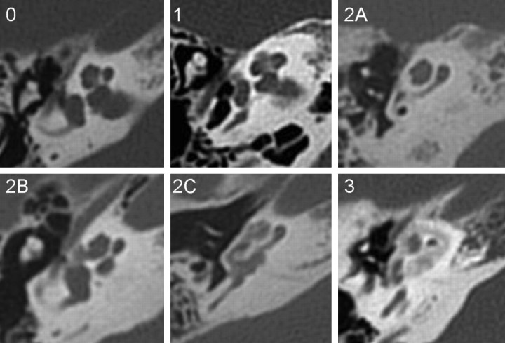
Fig 1.
Axial CT images of the petrous bone in patients with otosclerosis. Grade 0: normal. Grade 1: small lucent lesion at the fissula ante fenestram. Grade 2A: sclerosis and narrowing of the basal turn (also has spongiotic fenestral disease). Grade 2B: lucent lesion extending from the fissula ante fenestram to the middle turn of the cochlea. Grade 2C: patchy lucency around the lateral aspect of basal, middle, and apical turns of the cochlea, the medial aspect of the cochlea appears spared. Grade 3: severe, confluent lucency around the cochlea.

