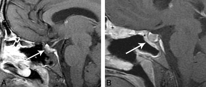
Fig 6.
A, Immediate postoperative sagittal T1-weighted postcontrast MR image with fat suppression shows no significant enhancement of the flap (white arrow). There is “C” shaped soft tissue in the defect that is presumably the flap (white arrow). B, Follow-up postoperative sagittal T1-weighted MR image postcontrast with fat suppression now shows an enhancing “C” nasoseptal flap (white arrow) in the surgical defect and increased enhancement to the tuberculum.

