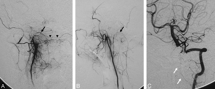
Fig 13.
Right ascending pharyngeal artery (A) and left vertebral artery (C) angiograms in the anteroposterior view and left ascending pharyngeal artery angiogram in the lateral view (B) show the anastomosis around the odontoid arch (arrowheads) with branches from the neuromeningeal trunks (black arrows) and the C3 segment of the left vertebral artery (white arrows).

