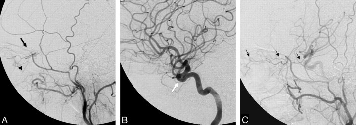
Fig 2.
Left ECA (A) and left ICA (B) angiograms (lateral view) demonstrate a meningo-ophthalmic artery arising from the left MMA, just before it crosses the sphenoid ridge on the lateral view (black arrow), contributing supply to the entire orbit with the absence of the ophthalmic artery from the ICA (white arrow). Note the choroidal blush (arrowhead). C, Right ECA angiogram of the same patient shows anastomosis between the MMA and the ophthalmic artery through the lacrimal system with retrograde filling of the ICA (thin black arrows).

