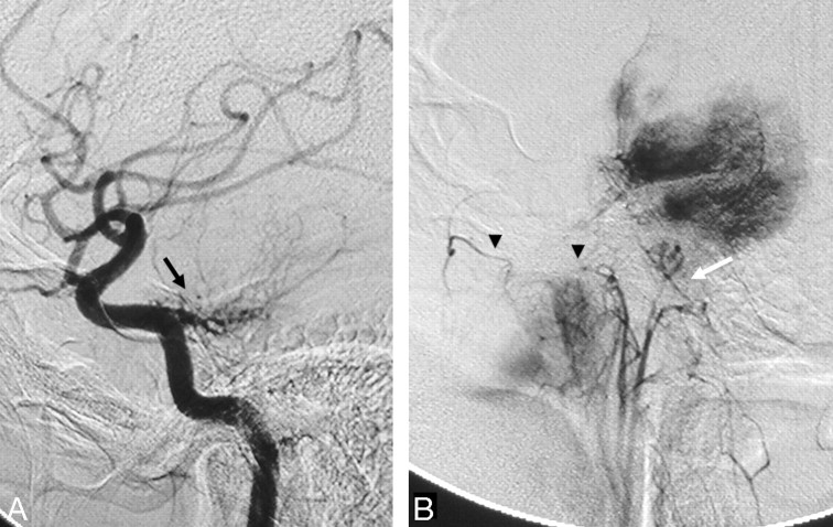
Fig 6.
Right ICA (A) and right ascending pharyngeal (B) angiograms in a lateral view reveal the typical “sunburst” appearance of a clival meningoma supplied by the meningohypophyseal trunk from the ICA (black arrow) and clival branches from the neuromeningeal trunk of the ascending pharyngeal artery (white arrow). Retrograde filling of the pterygovaginal artery of the distal IMA from the superior pharyngeal artery through the eustachian tube anastomotic circle is also seen (arrowheads).

