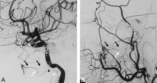
Fig 9.
Right ICA (A) and right ECA (B) angiograms in lateral view, posttransvenous coiling of a cavernous dural arteriovenous malformation, demonstrate the anastomosis between the vidian artery from the distal IMA to the vidian branch of the mandibular artery. The vidian artery has a characteristic horizontal course on the lateral view (black arrows), thus differentiating it from the more inferior course of the pterygovaginal artery (white arrows), which also anastomoses with a branch of the mandibular artery (black arrowhead) and the superior pharyngeal artery (white arrowhead) around the eustachian tube.

