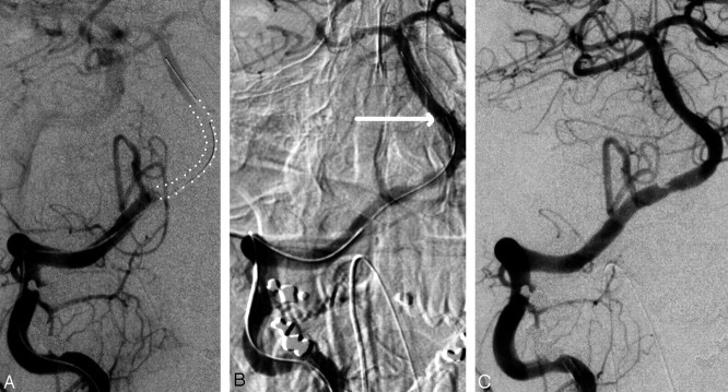
Fig 1.
A, Right vertebrobasilar arteriogram, dual-injection technique. Right VA occlusion distal to the posterior inferior cerebellar artery origin. A large vertebrobasilar thrombus is outlined by the dotted lines. B, Right VA angiogram, close-up view. The 4.5 × 37 mm Enterprise stent (Cordis) is partially unsheathed. Close inspection of the image reveals 3 small dots marking the distal end of the stent (arrow). The proximal end is still within its sheath, allowing recapture. Recanalization is already successful but has not yet reached its full extent. C, Right VA angiogram, 10 minutes after B was obtained. The stent is still partially deployed. Recanalization continues to improve (relative to A). After this run, the stent was recaptured and withdrawn.

