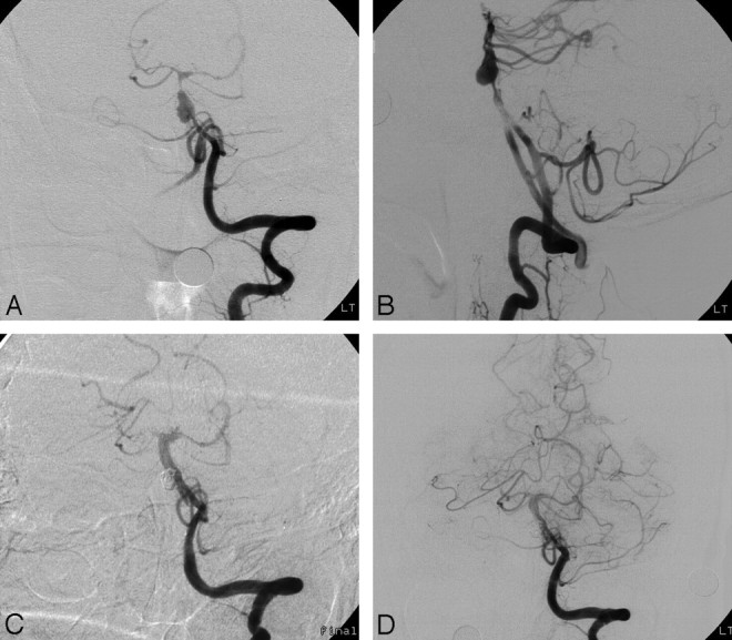
Fig 2.

Case 14 of an ruptured vertebrobasilar dissecting aneurysm. A and B, Anteroposterior and lateral angiograms of the left VA demonstrate a pearl-and-string sign with an approximately 5-mm pseudoaneurysm in the basilar artery. C, Anteroposterior angiogram of the left VA immediately after deployment of a balloon-expandable stent into the dissected basilar lesion and occlusion of pseudoaneurysm with 4 Guglielmi detachable coils. D, Follow-up anteroposterior angiogram of the left VA 11 days later shows good patency of the basilar artery with occlusion of the pseudoaneurysm.
