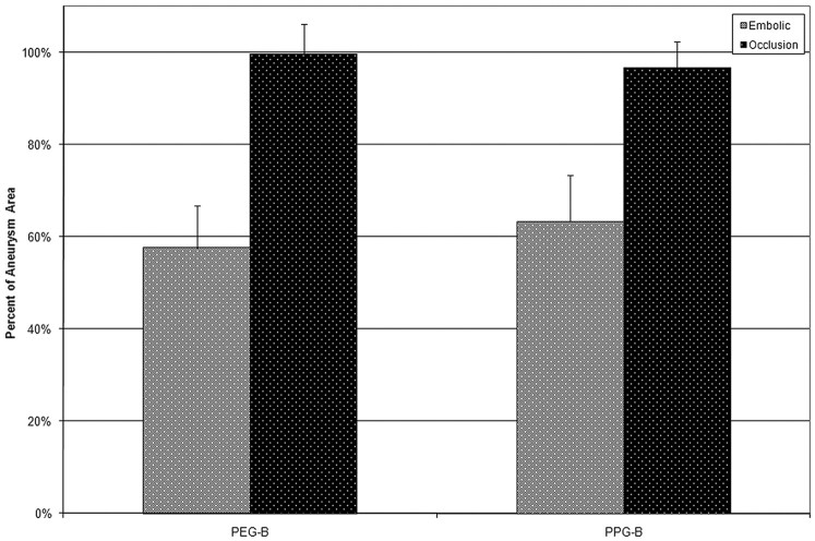
Fig 6.
Quantification of embolic and occluded areas from histologic sections. Because of the large amount of protrusion into the parent artery, the results from the PEG-I group are omitted from this figure. For both the PEG-B and PPG-B filaments, the average percentage of the aneurysm cavity occupied by embolic devices on the evaluated slides was approximately 60%. The average percentage of the aneurysm cavity that was occluded was approximately 100%.

