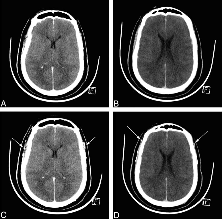
Fig 2.
CTA in BD before and 60 seconds after contrast medium injection. A and B, Unenhanced CT sections. C and D, Corresponding CT sections with identical window settings 60 seconds after contrast medium injection, demonstrating cerebral CT silence: absence of visualization of ICVs and cortical segments of the MCAs. Both superficial temporal arteries are opacified, indicating that contrast medium has been correctly injected (arrows).

