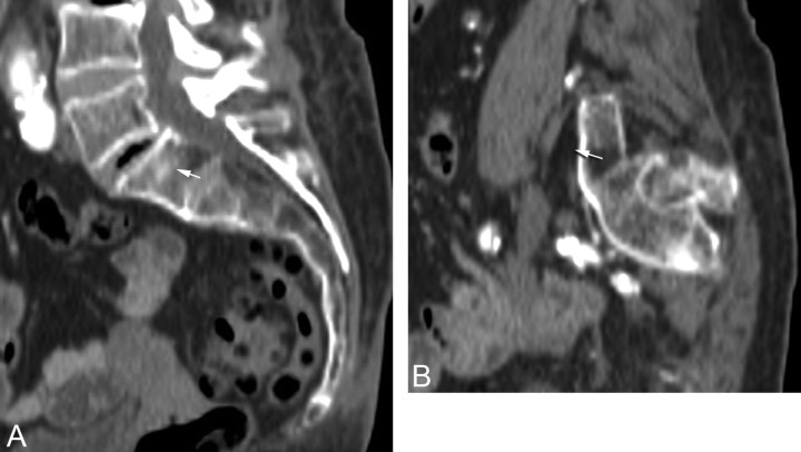
Fig 3.
Example of a potential breach of the anterior sacral cortex in an 88-year-old woman with lumbarization of S1. A, Midline sagittal reformatted image from a CT scan shows the target zone (arrow) within S1 as described in Fig 1. B, Left parasagittal reformatted image shows that needle placement (arrow) would be anterior to the sacral cortex.

