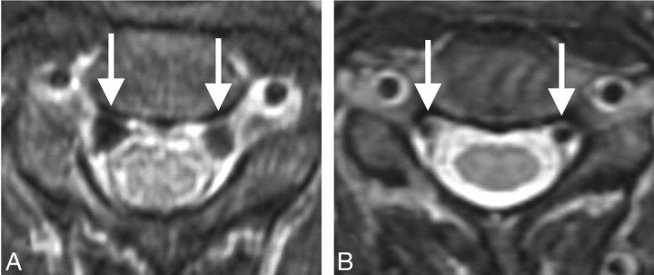
Fig 1.
Patient 10 had orthostatic headache. CSF pressure was negative, and brain MR imaging demonstrated diffuse pachymeningeal enhancement, tonsilar herniation, brain stem sagging, and subdural fluid collection. Spinal MR imaging showed distended epidural veins and epidural fluid collection at the cervical spine. The patient was successfully treated with epidural blood patch. A, MR image at the C2 level before treatment shows distention of the epidural veins (arrows). Collapsed dural sac appears as a hexagonal contour. B, Expansion of the dural sac and reduction of the venous size (arrows) are shown after treatment.

