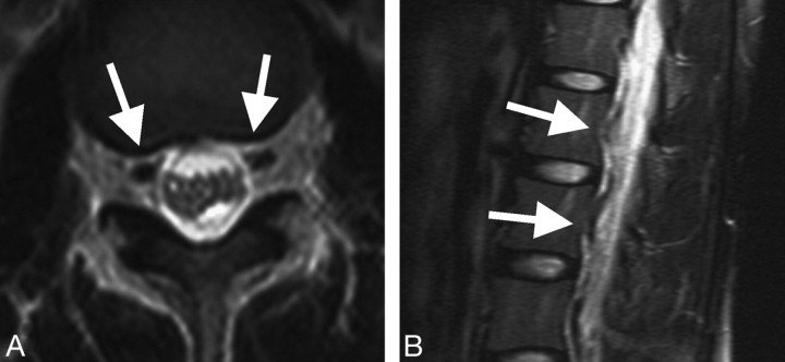
Fig 2.
Patient 4 presented with orthostatic headache and was diagnosed with SIH. Brain MR imaging demonstrated diffuse pachymeningeal enhancement, tonsillar herniation, sagging of the brain stem, and enlargement of the pituitary gland. Radioisotope cisternography revealed rapid excretion of tracer into the urine and CSF leakage at the lumbar spine. Thoracolumbar spinal MR imaging showed a prominent flow void. Her headache resolved after epidural blood patch treatment. A, Axial MR image shows prominent flow voids (arrows) in the bilateral anterolateral portions of the epidural space at the thoracolumbar region. B, Fat-saturated T2-weighted MR image at the thoracolumbar spine shows winding signal-intensity flow voids of the dilated epidural veins (arrows) behind the vertebral bodies on the parasagittal plane.

