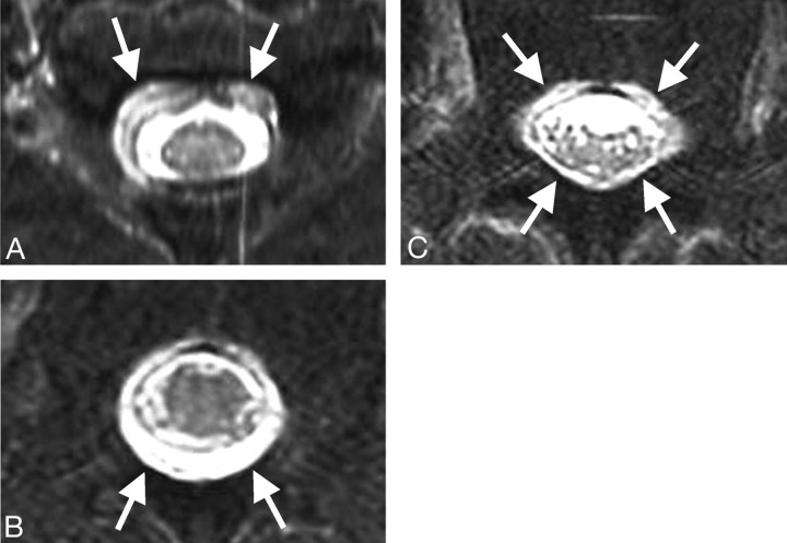
Fig 3.
Patient 1 had sudden onset of headache persisting for 1 month and was transferred to our institution with possible diagnosis of subarachnoid hemorrhage. Brain MR imaging revealed typical findings of SIH, including diffuse dural enhancement. Whole-spine MR imaging demonstrated epidural fluid collection. Epidural blood patch was effective, and the symptoms resolved. A–C, Axial fat-saturated T2-weighted MR images show fluid collection before treatment. Fluid collection (arrows) is visualized in the anterior epidural space at the cervical region (A), prominent in the posterior space at the thoracic region (B), and peripherally at the lumbar region (C).

