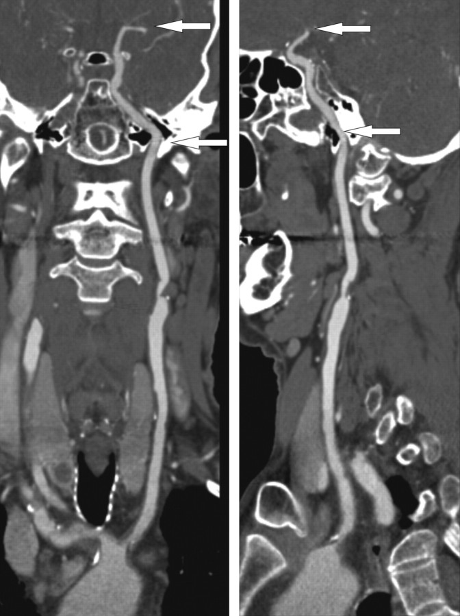
Fig 1.
A curved planar reformatted image from the aortic arch up to the top of the internal carotid artery. The arrows indicate the part of the internal carotid artery at which calcifications are segmented on axial sections. This part comprises the horizontal segment of the petrous internal carotid artery to the top of the carotid artery.

