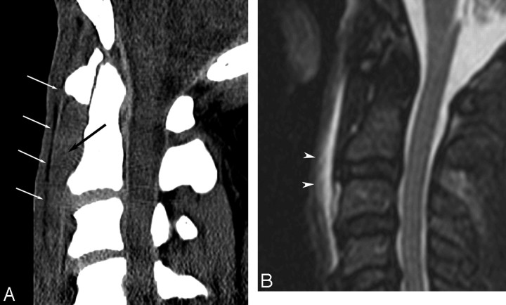
Fig 4.
A 43- year-old patient after a motor vehicle crash with no osseous injury. A, Midsagittal MDCT image of the cervical spine demonstrates abnormal high attenuation (black arrow) anteriorly displacing the retropharyngeal fat plane (white arrows). B, Short τ inversion recovery MR image obtained the same day shows extensive PVST edema and/or hematoma in this region (arrowheads).

