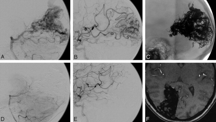
Fig 2.
A 29-year-old female patient with an SM grade IV AVM in the right occipital lobe. A, Vertebral angiogram (lateral view) shows arterial supply from multiple feeders arising mainly from the right posterior artery with superficial venous drainage. B, Right internal carotid artery angiogram (lateral view) shows additional arterial supply to the AVM from feeders of the middle cerebral artery. C, Unobstructed image illustrates a solid Onyx cast after the embolization. D and E, Control right vertebral and carotid artery angiograms (lateral view) at 6 months after embolization illustrate persistence of complete occlusion with no evidence of recanalization. F, MR image (time-of-flight, axial) at 6 months after embolization reveals complete occlusion of the AVM without evidence of reperfusion.

