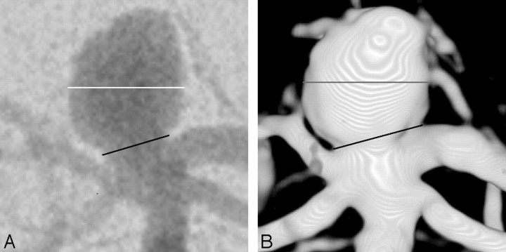
Fig 1.
Images from a patient with a basilar tip aneurysm. A, Anteroposterior (AP) 2D DSA shows the measurement of dome diameter (white line) and neck width (black line). Dome-to-neck ratio for this 2D DSA image is 1.7. B, AP 3DRA from the same patient as in A shows dome diameter (gray line) and neck width (black line). Dome-to-neck ratio for this 3DRA image is 1.3.

