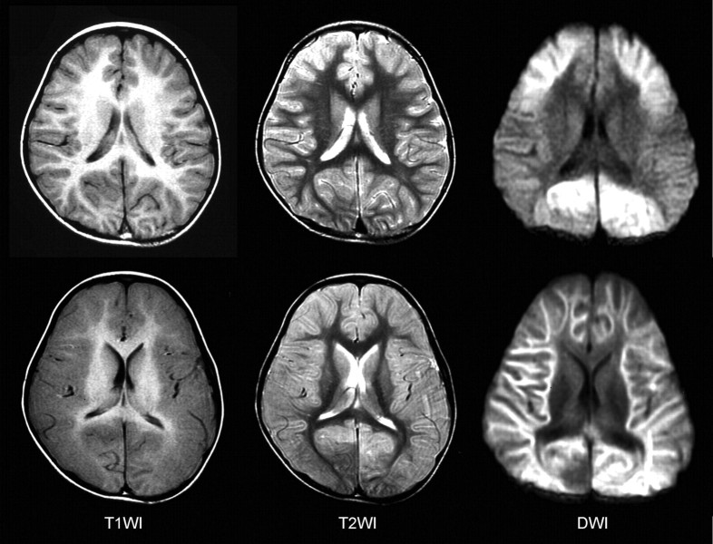
Fig 1.
MR imaging findings of a patient with diffuse lesions. Top: The day after the onset, T1-weighted (T1WI) images show mild thickening of the cortex and T2-weighted (T2WI) images reveal mildly increased intensities in the cortex of the bilateral frontal lobes. Reduced diffusivity is observed in the bilateral frontal and occipital regions on DWIs (frontal occipital lesions). Bottom: Five days after the onset, T1WI and T2WI images demonstrate marked edematous changes in the entire cortex. Reduced diffusivity is observed in the entire subcortical white matter on DWIs.

