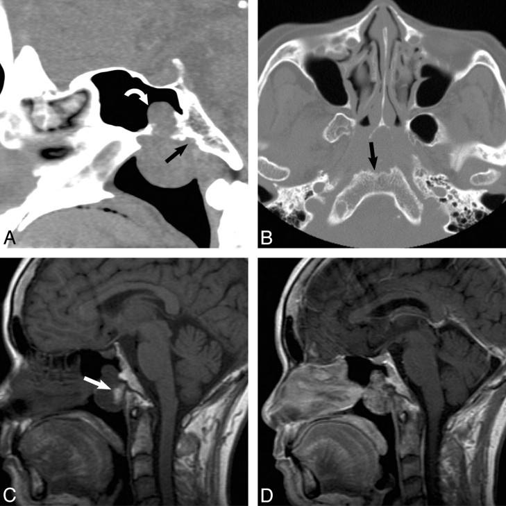
Fig 1.
A, A 65-year-old woman presented with nasal obstruction. Findings show atypical CT appearance of a pathology-proved extraosseous nasopharyngeal chordoma. Sagittal noncontrast CT scan shows a lobulated mass centered in the nasopharynx with superior extension into the sphenoid sinus (curved arrow) and focal erosion of the anterior clivus (black arrow). B, Axial bone CT scan shows a lobulated soft-tissue mass centered in the nasopharynx, with scalloping of the anterior margin of the clivus. Note the lytic appearance with a sclerotic margin (black arrow). C, Sagittal T1-weighted MR image shows a heterogeneous nasopharyngeal mass with a focal area of T1 hyperintensity (arrow) thought to represent blood products or proteinaceous debris. D, Sagittal T1-weighted MR image with contrast shows heterogeneous enhancement of the mass, which is an enhancement pattern seen in both extraosseous and typical skull base chordomas.

