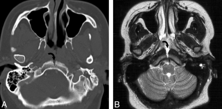
Fig 4.
A, A 32-year-old woman presented with a mass in the nasopharynx. MR imaging shows a midline sinus tract extending into the clivus of a pathologically proved extraosseous nasopharyngeal chordoma. Axial bone CT scan shows a midline tract (curved arrow) representing the extension of the extraosseous chordoma into the medial basal canal. B, Axial T2-weighted MR image shows the hyperintense midline sinus tract (curved arrow) extending posteriorly from the nasopharyngeal mass. Note fluid in the left mastoid air cells secondary to obstruction of the eustachian tube due to a nasopharyngeal chordoma.

