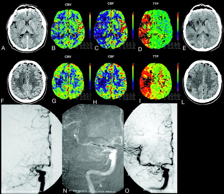
Fig 2.

A 35-year-old man with left hemiplegia, imaged 3 hours after symptom onset. NCCT, CBV, CBF, and TTP maps, delayed NCCT (A−E, first level; F−L, second level); and IAT (M−O). NCCT shows mild hypoattenuation of the right lenticular nucleus. PCT color maps show small multiple areas with severely reduced CBV (CBV ratio <33%) and CBF (<11.5 mL/100 g/min) in the anterior third of the right lenticular nucleus; external capsule; and fronto-opercular, insular, posterior temporal and parietal cortices, corresponding to the infarct core (solid line). The infarct core is surrounded by a large perfusion deficit in the right MCA cortical territory, characterized by reduced CBF (color-coded blue) and increased TTP (color-coded red), indicating a TTP-CBV mismatch, which corresponds to the ischemic penumbra. IADSA, performed after PCT, shows proximal right M1 occlusion (M), with poor collateral leptomeningeals. The patient underwent intra-arterial thrombolysis with injection of rtPA and mechanical clot manipulation, followed by percutaneous transluminal angioplasty, with complete right MCA recanalization (N). A mild residual M1 stenosis is identifiable at the end of the procedure (O). Follow-up CT scans (E and L), obtained 2 days after stroke, show small multiple infarcts in the right caudate and anterior third of the lenticular nucleus; insular cortex; and temporal, frontal, and parietal cortices, which correspond to the infarct core, with consequent recovery of a large portion of the mismatch area. At 3 months, the patient was independent (mRS score, 1).
