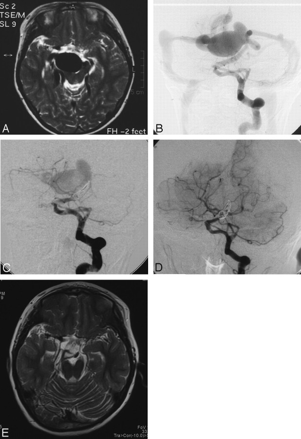
Fig 3.
Patient 3. A, T2-weighted MR imaging of the head at the level of the midbrain shows giant flow-void signal intensity in the interpeduncular cistern. B, Anteroposterior view of the left vertebral artery injection demonstrates that the fistula is supplied by the primitive trigeminal artery and drained into the basal vein of Rosenthal, with an associated venous varix. C, After coil embolization, anteroposterior view of the left vertebral artery injection demonstrates incomplete disconnection of the fistula by the coils. D, Follow-up angiogram obtained at 7 months shows spontaneous occlusion of the fistula with preserved patency of the basilar artery. E, Disappearance of the venous varix on follow-up T2-weighted MR imaging.

