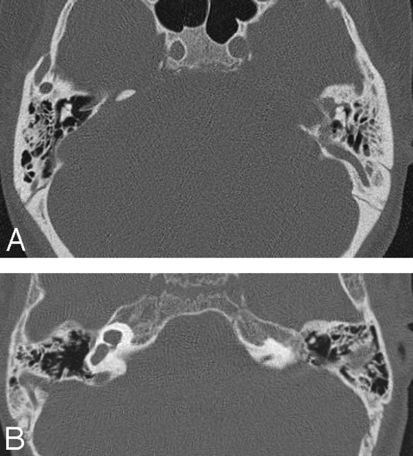
Fig 1.
A, Axial CT scan of the temporal bones of patient 1 shows bilateral absence of total inner ear structures with aplasia of the otic capsules bilaterally. Note a large emissary vein on the left. B, CT scan of patient 4 reveals unilateral CLA on the left with a type 1 incomplete cochlear partition on the right. The petrous bone and the otic capsule are hypoplastic on the left.

