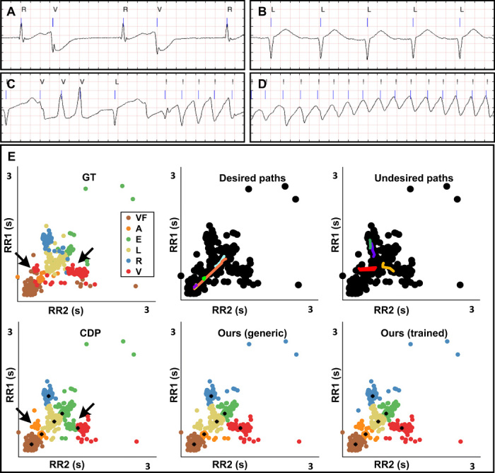
Fig. 6. Recognition of different heart rhythms.

(A to D) Fragments of an ECG signal from a patient with several types of arrhythmia. Blue lines indicate a beat (systole). Cardiologists labeled six different types of beats “A, V, E, R, L, and !,” associated with different rhythms. Atrial/ventricular/ectopic beats “A/V/E” are extrasystoles close to the previous beat that can induce a pause in the rhythm (A). Right bundle branch block “R”: beat appearing in this patient between two V. Left bundle branch block “L” (B): normal beat type for this patient (similar RR1 and RR2 varying linearly in tachycardia and bradycardia). Runs of ventricular extrasystoles not followed by long pause are also reported (C). Ventricular flutter “!,” later indicated as “VF,” a critical condition characterized by extremely fast contractions (D). (E) Heartbeats represented as points, described according to their distance from the previous and to the next beat (RR1 and RR2). Our method recognized ventricular extrasystoles (red), which are otherwise mixed with L beats by CDP. The trained version of the algorithm further allowed identification of a group composed of mixed atrial and ventricular extrasystoles (orange).
