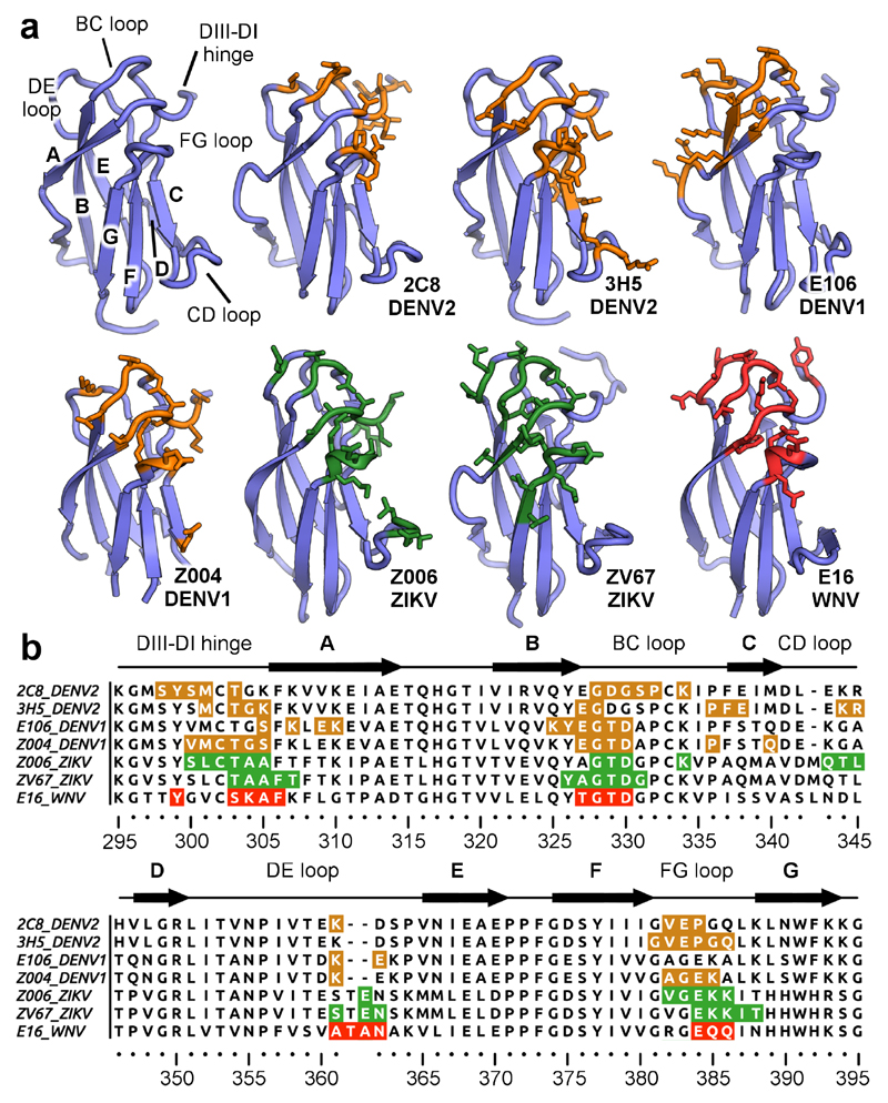
Figure 5. Epitope recognition by 3H5 and 2C8.
a, EDIII epitopes recognized by lateral ridge antibodies directed against different flaviviruses. The antibody designations and targeted viruses are indicated. The general architecture of EDIII is shown in the top left and important regions are labelled. Residues engaged by antibodies on dengue virus EDIIIs are colored in orange, ZIKV EDIIIs in green and WNV EDIIIs in red. All contacted residues are rendered as sticks. b, Sequence alignment of DENV, ZIKV, and WNV EDIIIs with highlighted antibody epitopes (coloring as above, antibodies and viruses indicated on the left of sequences). EDIII regions are labelled above the sequences.

