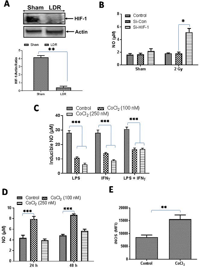
Figure 1. Dual role of HIF-1 in macrophage retuning.
A) RT5 mice (25 weeks of age) were treated twice with 2 Gy irradiation and were analyzed for the expresion of HIF-1 as an indicator of tumor hypoxia. A representative blot from several tumor lysate repeats with similar outcomes is shown. β-actin was used a loading control. Densitometric analysis of all the western blots were quantified byImageJ software and the mean densitometry values were plotted in terms of relative protein expression. (B)HIF-1 proteins were depleted in RAW macrophages by siRNA-based methods and the macrophages were then irradiated with a dose of 2 Gy and the impact of this treatment was analyzed on the level of NO in the cell culture supernatants. (C) RAW macrophages treated with various doses of CoCl2 (to stabilize the expression of HIF-1) were stimulated with Th1 effectors (i.e. LPS and IFNγ) and NO levels were quantified 24h post treatment by using a Giess reagent method. (D) Naive RAW macrophages were treated with CoCl2 and the impact of treatment on the levels of NO were analyzed. Shown is the mean level (μM) of NO ± SEM from three independent experiments. (E) Expression of iNOS in CD11b+/Gr-1(−) RT5 tumor macrophages under the influence of CoCl2 treatment was analyzed. Statistical analyses were conducted using either Student’s t tests, 1 way or 2 way ANOVA, followed by Bonferroni post-test (*P < 0.05; **P < 0.01; ***P < 0.001). All the statistical analyses was carried out with GraphPad Prism Version 7.0 software.

