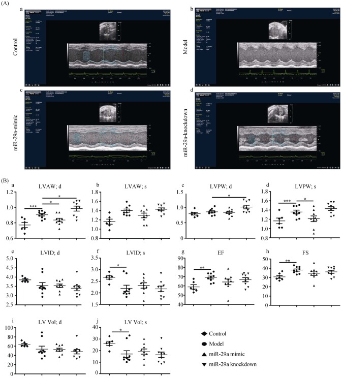
Figure 1. Overexpression of miR-29a inhibits left ventricular remodeling by using ultrasound assessment.
(A): Representative M-mode echocardiography images; and (B): echocardiography parameters were detected in mice among four groups (a. LVAW; d. the thickness of the LVAW at the end-diastolic; b. LVAW; s. the thickness of the LVAW at the end-systolic; c. LVPW; d. the thickness of the LVPW at the end-diastolic; d. LVPW; s. the thickness of the LVPW at the end-systolic; e. LVID; d. the left ventricular interior diameter at the end-diastolic; f. LVID; s. the LVID at the end-systolic; g. EF%: LV ejection fraction; h. FS%: LV fractional shortening; i. LV Vol; d. LV volume at the end-diastolic; and j. LV Vol; s. LV volume at the end-systolic.). Data are indicated as the means ± S.E. *P < 0.05, **P < 0.01, ***P < 0.001. EF: ejection fraction; FS: fractional shortening; LV: left ventricular; LVAW: left ventricular anterior wall; LVID: left ventricular inner dimension; LVPW: left ventricular posterior wall.

