Figure 2.
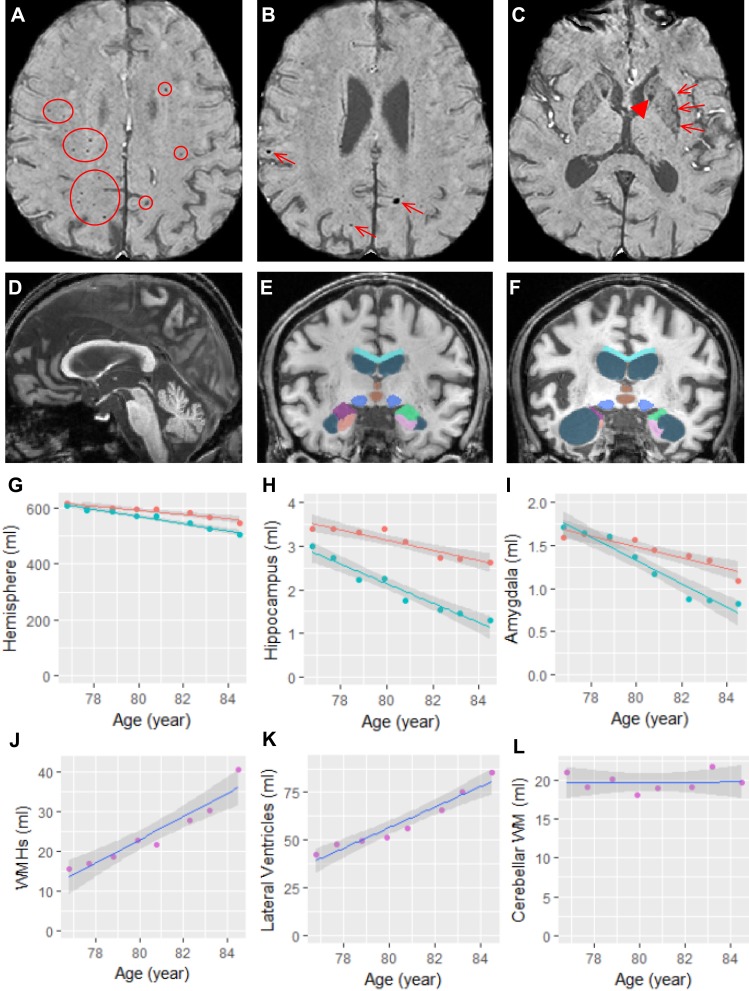
Structural changes in the brain. (A–C) The SWI scan acquired at age 76 shows cerebral microbleeds in the white matter and all four lobes (circled in (A) and arrows in (B)) as well as symmetric iron depositions in the caudate nuclei (arrowhead in (C)) and putamen (arrows in (C)). (D) The midsagittal slice of his T1 scan acquired at age 84 shows the relatively reserved the corpus callosum, brainstem, and cerebellum. (E and F) Segmentation of the corpus callosum (cyan), lateral ventricles (steel blue), third ventricle (brown), midbrain (blue), left amygdala (lime green), right amygdala (purple), left hippocampus (pink), and right hippocampus (salmon) on MRI scans acquired at age 76 (E) and age 84 (F). (G-L) Volumetric changes in left (red) and right (turquoise) hemisphere (G), left and right hippocampus (H), left and right amygdala (I), white matter hyperintensities (WMHs) (J), lateral ventricles (K), and cerebellar white matter (WM) (L).

