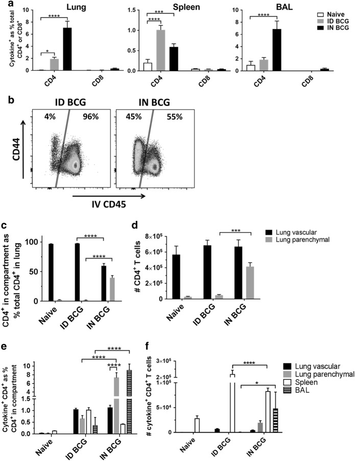
Fig. 2.
Mucosal BCG recruits CD4+ T cells to the lung parenchyma and induces greater frequency of antigen-specific CD4+ T cells in the lung parenchyma and BAL. Six weeks after immunisation with BCG via IN or ID route, intravascular staining and ICS identified populations of lung parenchymal and lung vascular antigen- (PPD-T)-specific (cytokine+) CD4+ or CD8+ T cells producing IFN-γ, TNF-α or IL-2 alone or in combination. a Frequency of BCG-induced antigen-specific cytokine+ CD4+ and CD8+ T cells in the lung, spleen and BAL. b Representative flow cytometry plots showing the proportion of lung parenchymal and lung vascular CD4+ T cells in the lungs of ID- or IN-immunised mice following intravascular anti-CD45 staining. c Frequency of lung parenchymal and lung vascular CD4+ T cells as a % of total CD4+ T cells isolated from the lung. d Number of CD4+ T cells in the lung parenchymal and lung vascular compartments. e Frequency of BCG-induced antigen-specific cytokine+ CD4+ T cells. f Number of BCG-induced antigen-specific cytokine+ CD4+ T cells. For a, c–f bars represent mean ± SEM (n = 6). Two-way ANOVA with Sidak’s post-test (a, c, d) or Tukey’s post-test (e, f); ****P < 0.0001, ***P < 0.001. Data are representative of one of two independent experiments

