Fig. 3.
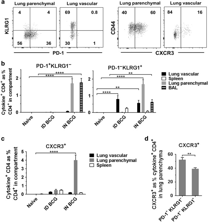
Mucosal, but not ID BCG, induces PD-1+ KLRG1− CD4+ T cells in the lung parenchyma and BAL. Six weeks after immunisation with BCG via IN or ID route, intravascular staining and ICS identified antigen-specific (cytokine+) PD-1+ KLRG1− and PD-1− KLRG1+ CD4+ T cells. a Representative plots from IN-immunised mice, pre-gated on cytokine+ CD4+ T cells, showing surface staining for PD-1, KLRG1 and CXCR3 in the lung parenchyma and lung vasculature. b Frequency of cytokine+ CD4+ T cells expressing a PD-1+ KLRG1− or PD-1− KLRG1+ phenotype. c Frequency of cytokine+ CD4+ T cells expressing CXCR3. d Proportion of lung parenchymal cytokine+ CD4+ T cells expressing CXCR3 in PD-1/KLRG1 subsets in mice after mucosal BCG vaccination. For b–d bars represent mean ± SEM (n = 6). Two-way ANOVA with Tukey’s post-test (b) or Sidak’s post-test (c); ****P < 0.0001, ***P < 0.001, **P < 0.01. Unpaired t-test (d); **P < 0.01. Data are representative of one of two independent experiments

