
Fig. 4.
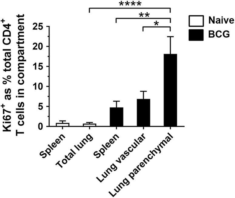
Lung parenchymal CD4+ T cells have greater proliferative capacity than lung vascular or splenic CD4+ T cells following mucosal BCG. Mice were vaccinated with BCG IN 6 weeks prior to intravascular staining. Purified populations of lung parenchymal, lung vascular and splenic CD4+ T cells were obtained by sorting. Naïve CD4+ lung and spleen T cells were sorted without intravascular staining. Cells were cultured for 3 days with PPD-T before proliferating cells were identified with Ki67 staining. To account for non-specific expression of Ki67, unstimulated values were subtracted from stimulated. Graph shows frequency of Ki67+ CD4+ T cells in the total lung and spleen of naïve mice and the lung parenchyma, lung vasculature and spleen of BCG-immunised mice. Bars represent mean ± SEM (n = 6). One-way ANOVA with Tukey’s post-test, ****P < 0.0001; **Ρ < 0.01, *Ρ < 0.05. Data are pooled from three independent experiments