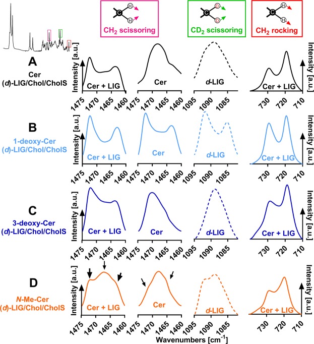
Figure 5.
Lateral lipid packing in the SC lipid model containing either Cer (panel A), or 1-deoxy-Cer (panel B), or 3-deoxy-Cer (panel C), or N-Me-Cer (panel D), along with (d)-LIG, Chol, and CholS. First graphs in each panel show the representative infrared spectra of the methylene scissoring vibrations of the unlabeled lipid mixtures at 32 °C. Second and third graphs in each panel show the CH2 scissoring vibration (mainly from Cer) and CD2 scissoring vibrations (d-LIG; dashed line), respectively, of the lipid mixtures with d-LIG at 32 °C. The fourth graphs in each panel show the representative infrared spectra of the methylene rocking vibrations of the unlabeled mixtures at 32 °C. A presence of a CH2/CD2 doublet in the spectrum is indicative of an orthorhombic packing. Intensity is given in arbitrary units (a. u.).

