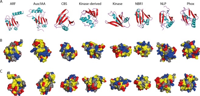
Figure 6.
Representative homology model structures of PB1 domain from one member from each family in Arabidopsis. The identifiers are: ARF5 (PDB: 4CHK), IAA17 (PDB: 2MUK), At2G36500 (CBS), At1G04700 (Kinase), At2G01190 (Kinase-derived), NBR1, NLP9 and Phox2. (A) Secondary structures shown in various colours: α-helices in ‘Cyan’; β-sheets in ‘red’ and turns in ‘purple’. Surface representation of the positive and negative faces shown in (B,C) respectively. Hydrophobic amino acids ‘AGVILFMP’ are in ‘Yellow’; Polar residues ‘NQTSCYW’ are in ‘Grey’; Positively charged ‘RKH’ are in ‘Blue’ and Negatively charged ‘DE’ are in ‘Red’.

