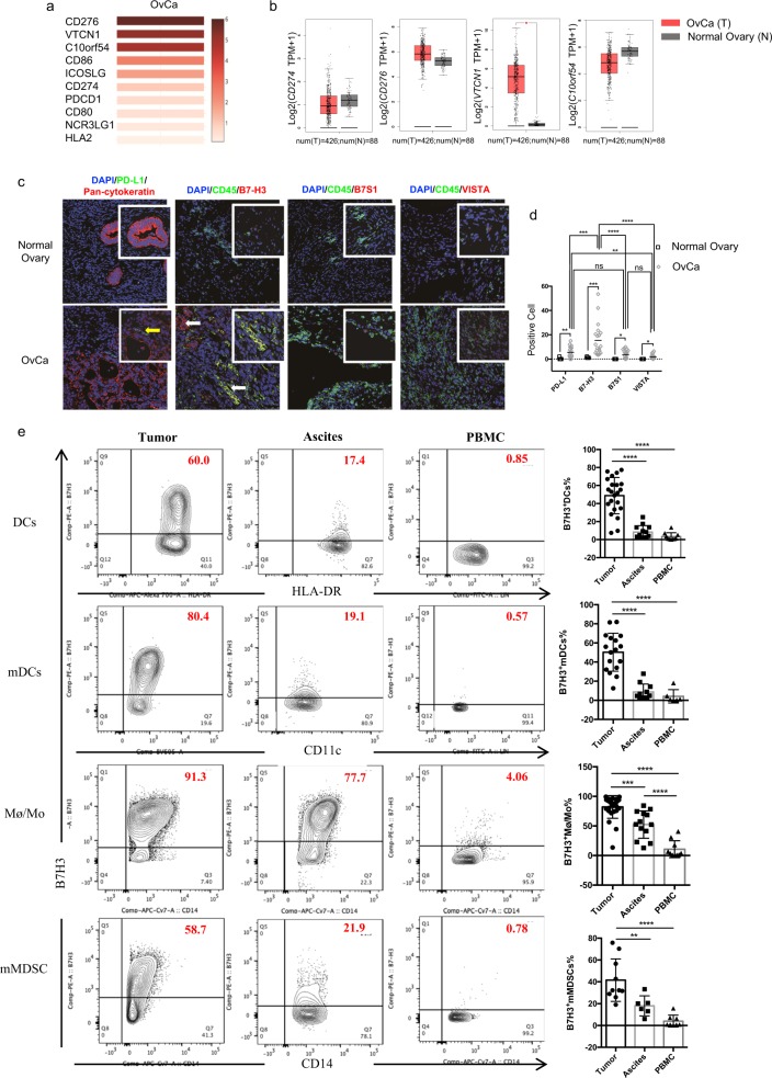
Fig. 1.
B7-H3, but not PD-L1, is robustly expressed in human OvCa. a Heatmap analysis of the mRNA expression of B7 family genes in OvCa tumors shown as scaled log2-fold changes (GEPIA data). b The mRNA expression levels of CD274, CD276, VTCN1, and C10orf54 in human OvCa tumor tissues and normal ovarian tissues. The data were derived from the TCGA database, and are shown on a log2(TPM + 1) scale. TPM: transcripts per million. The P-value cutoff was 0.01. c Representative immunofluorescence images of PD-L1, B7-H3, B7S1, and VISTA in human OvCa and normal ovarian tissues. Original magnification ×40; scale bar, 50 μm. Inset original magnification ×100; scale bar, 25 μm. d Quantification of PD-L1+, B7-H3+, B7S1+, and VISTA+ cells in tumor and normal tissues (cell numbers were quantified in images of a ×100 field). Each dot represents the data from one patient. One-way ANOVA followed by Tukey’s multiple comparison test. e Representative figures and summarized data showing the percentages of B7-H3-positive mDCs, DCs, Mø/Mo, and mMDSCs in tumors, ascites, and PBMCs from OvCa patients. One-way ANOVA followed by Tukey’s multiple comparison test. The data are presented as the mean ± SEM; ns P > 0.05, *P < 0.05, **P < 0.01, ***P < 0.0001, and ****P < 0.00001

