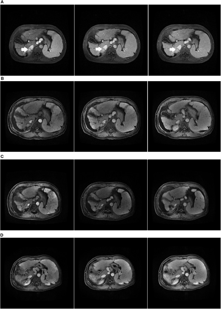
Figure 6.
Child-Pugh C HCC patient who received CK-SBRT assessed by MRI. (A) The initial abdominal MRI scan with the primary HCC indicated by the arrow. (B) MRI scan of 3 months after SBRT. (C) MRI scan of 24 months after SBRT. The lesion in the liver disappeared. (D) MRI scan of 42 months after SBRT.

