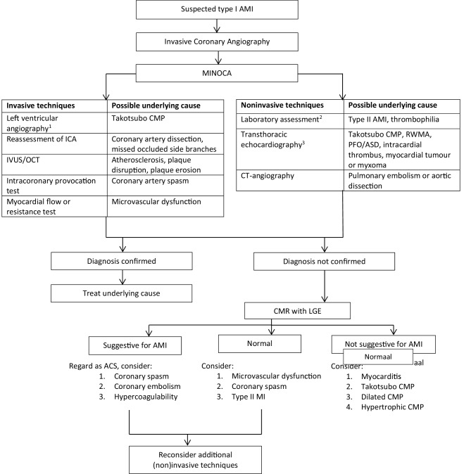
Fig. 2.
Proposal for a diagnostic algorithm in patients with myocardial ischaemia with non-obstructive coronary arteries. aUnless renal function <35 ml/min per 1.73 m2. bHaemoglobin, C‑reactive protein, leucocytes, oxygen saturation, D‑dimers, (NT-pro) brain natriuretic peptide. c Within 48 h. AMI acute myocardial infarction, MINOCA myocardial ischaemia with non-obstructive coronary arteries, ICA invasive coronary angiography, CMP cardiomyopathy, IVUS intravascular ultrasound, OCT optical coherence tomography, RWMA regional wall motion abnormalities, PFO patent foramen ovale, ASD atrial septal defect, CT computed tomography, CMR cardiac magnetic resonance imaging, LGE late gadolinium enhancement. ACS acute coronary syndrome

