Fig. 5.
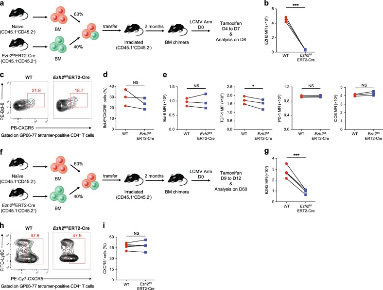
EZH2 is not essential for the late differentiation and maintenance of virus-specific TFH cells during an acute viral infection. a Setup of the BM chimera experiment. Irradiated CD45.1+ WT recipients underwent adoptive transfer of CD45.1+ WT BM cells (60%) and CD45.2+ Ezh2fl/flERT2-Cre BM cells (40%). Two months after reconstitution, the recipients were infected with LCMV Armstrong, followed by the administration of tamoxifen from days 4 to 7 after infection and an analysis on day 8. b Expression of EZH2 in virus-specific Bcl6hiCXCR5+ TFH cells from the BM chimeric WT and Ezh2fl/flERT2-Cre mice shown in a. c Flow cytometry analysis of GP66–77 tetramer-positive CD4+ T cells in the spleens of the chimeras described in a on day 8 after the LCMV Armstrong infection. Numbers adjacent to outlined areas indicate the percentage of Bcl-6hiCXCR5+ TFH cells, which were summarized in d. e Levels of Bcl-6, TCF-1, PD-1 and ICOS in the Bcl-6hiCXCR5+ TFH cells shown in c. f The BM chimeras described in a were infected with LCMV Armstrong, treated with tamoxifen on days 9 to 12 after infection and analyzed on day 60. g Quantification of EZH2 expression in virus-specific CXCR5+ TFH cells originating from BM chimeric WT and Ezh2fl/flERT2-Cre mice shown in f. h Flow cytometry of GP66–77 tetramer-positive CD4+ T cells in the spleens of the chimeras described in f on day 60 after the LCMV Armstrong infection. Numbers adjacent to outlined areas indicate the percentage of CXCR5+ TFH cells, which were summarized in i. NS not significant; *P < 0.05 and ***P < 0.001 (paired two-tailed t-test (b, d, e, g and i)). The data are representative of two independent experiments with at least three mice (b, d, e, g, h and i) per group

