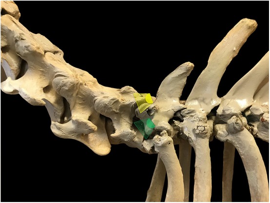
Figure 1.

Ultrasound probe positioning and orientation to visualize C8 ramus ventralis in a longitudinal section in horses. The probe is initially oriented perpendicular to the vertebral axis to obtain a reference image of the 2 articular processes and the joint space (yellow scan field) then glided ventro-caudally with an approximate 20° angle from its initial position (counter-clockwise rotation on the left side of the neck, green scan field).
