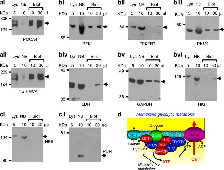
Fig. 4. Glycolytic enzymes are associated with plasma membrane proteins in Mia PaCa-2 cells.
Mia PaCa-2 cells were incubated with sulfo-NHS-SS-Biotin, which binds to primary amines on cell surface/transmembrane proteins. These biotinylated proteins were separated from non-biotinylated proteins using NeutrAvidinTM agarose beads and eluted using dithiothreitol. Whole-cell lysates (Lys), non-biotinylated (NB) and biotinylated (Biot) fractions were separated by SDS-PAGE and western blotted for the membrane proteins, PMCA4 (ai) and non-specific (NS) PMCA (aii), the glycolytic enzymes, phosphofructokinase-1 (PFK1, bi), phosphofructokinase fructose bisphosphatase-3 (PFKFB3, bii) and pyruvate kinase-M2 (PKM2, biii), lactate dehydrogenase (LDH, biv), glyceraldehyde phosphate dehydrogenase (GAPDH, bv), hexokinase-I (HKI, bvi) and the mitochondria-associated proteins hexokinase-II (HKII, ci) and pyruvate dehydrogenase (PDH, cii). d Cartoon depicting a complex of key glycolytic enzymes associated with the plasma membrane (membrane glycolytic metabolon) in close proximity to glucose transporters (GLUT), lactate transporters (MCT) and the PMCA.

