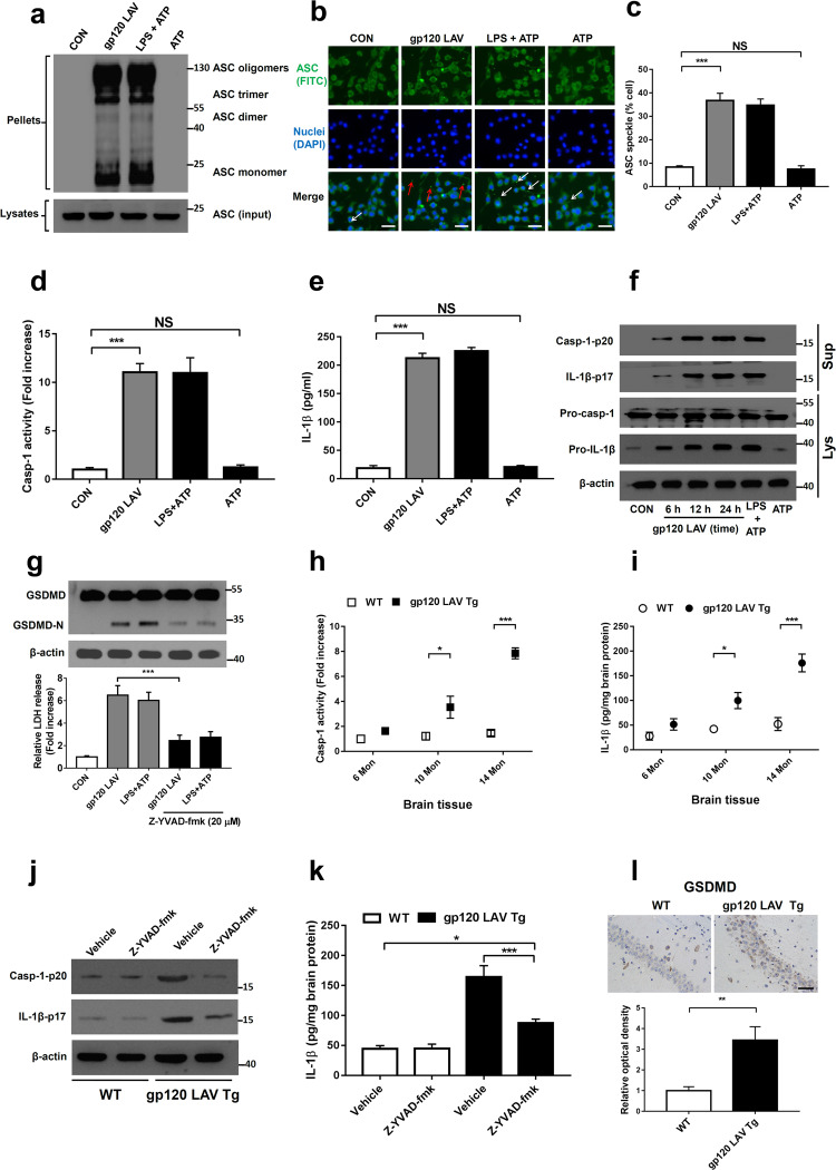
Fig. 1.
HIV-1 gp120 LAV induces inflammasome activation in vitro and in vivo. a–c Western blot of ASC in crosslinked pellets (upper panel) and cell lysates (lower panel) (a) and fluorescence microscopy of ASC specks (b) of BV2 cells treated with control, gp120 LAV (0.5 μg/ml), LPS + ATP or ATP. The arrows indicate speck formation, scale bar: 20 μm. c The percentages of ASC specks in BV2 cells. Cells primed with LPS and stimulated with ATP (LPS + ATP) served as a positive control. d, e Caspase-1 activity (d) and IL-1β release (e) in supernatants from BV2 cells treated with control, gp120 LAV (0.5 μg/ml), LPS + ATP or ATP. The data in (d) are expressed as the ratio of the absorbance determined from the treated cells to that of the control. f Western blots of activated caspase-1 (Casp-1-p20) and cleaved IL-1β (IL-1β-p17) in culture supernatants (Sup) from BV2 cells treated with gp120 LAV (0.5 μg/ml) at different times. Pro-casp-1 and pro-IL-1β in cell lysates (Lys) were also detected. g GSDMD immunoblot analysis (upper panel) and LDH release (lower panel) of BV2 cells treated with gp120 LAV (0.5 μg/ml) in the absence or presence of Z-YVAD-fmk (20 μM). LDH data are expressed as the ratio of the absorbance determined from the treated cells to that of the control. h, i Caspase-1 activity (h) and IL-1β level (i) in brain tissues of WT and gp120 LAV transgenic (Tg) mice at different ages (6, 10, 14 months, Mon, n = 5). j, k WT (n = 5) and gp120 LAV Tg mice (n = 5) treated with or without Z-YVAD-fmk (10 mg/kg body weight) for 30 days. Then, all mice were euthanized by CO2 inhalation, and the brain tissue was harvested and homogenized. j Immunoblots of Casp-1-p20 and IL-1β-p17 and k ELISA of IL-1β in brain tissue homogenates. l Representative images of immunolabeled GSDMD from the hippocampal section of WT and gp120 LAV Tg mice (n = 3, upper panel); optical density was analyzed quantitatively (n = 6, lower panel), Scale bar: 50 μm. The data are displayed as the mean ± SEM (n = 5, c, d, e, g, h, i, k; n = 6, l) from three independent experiments. The western blot, fluorescence and immunohistochemical results are representative of three independent experiments (a, b, f, g, j, l). *P < 0.05, **P < 0.01, ***P < 0.001; NS, no significance

