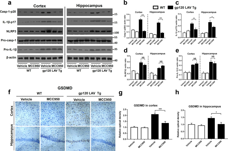
Fig. 6.
Effect of MCC950 on NLRP3 inflammasome activation in gp120 LAV Tg mice. a–e Western blots (a) and densitometric analysis (b–e) of activated caspase-1 (Casp1-p20, b), cleaved IL-1β (IL-1β-p17, c), NLRP3 (d), and pro-IL-1β (e) in the cortex and hippocampus of WT (n = 5) and gp120 LAV Tg (n = 5) mice administered vehicle (PBS, 80 days) or MCC950 (10 mg/kg BW, dissolved in PBS, 80 days). f Brain sections of the cortex (upper panel) and hippocampus (lower panel) were immunostained with GSDMD, scale bar: 100 μM. g, h Images of immunolabeled GSDMD were analyzed quantitatively using ImageJ software. The Western blots and immunohistochemical results are representative of two independent experiments (a, f); data are displayed as the mean ± SEM (n = 5, b–e) and (n = 6, g, h) from two independent experiments. *P < 0.05, **P < 0.01, ***P < 0.001; NS, no significance

