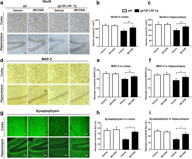
Fig. 8.
Effect of MCC950 on neuronal damage in gp120 LAV Tg mice. a, d Representative images of the cortex (upper panels) and hippocampus (lower panels) immunolabeled with NeuN (a), MAP-2 (d), and synaptophysin (g) from WT (n = 3) and gp120 LAV Tg (n = 3) mice administered vehicle (PBS, 80 days) or MCC950 (10 mg/kg BW, dissolved in PBS, 80 days). b, c, e, f, h, i Quantification of microscopy data obtained in the cortex and hippocampus: quantification of NeuN-positive cells (counts/0.1 mm2) in the cortex (b) and hippocampus (c); MAP-2 and synaptophysin represented by the percentage of positive neuropils. Scale bar: 100 μm. The immunohistochemical staining results are representative of two independent experiments (a, d, g). The data are displayed as the mean ± SEM (n = 6) from two independent experiments (b, c, e, f, h, i). *P < 0.05, **P < 0.01

