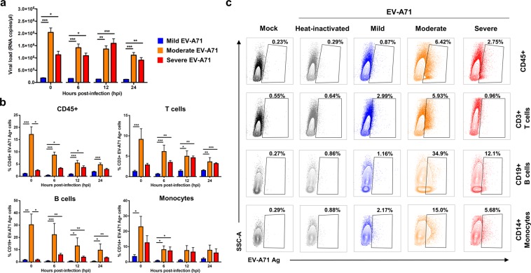
Figure 2.
EV-A71 infection in primary human peripheral blood mononuclear cells (PBMCs). Human primary PBMCs (n = 5–9) were infected with mild, moderate, severe and heat-inactivated EV-A71 isolates at MOI 5, and harvested at 0, 6, 12 and 24 hpi for viral load quantification and flow cytometry. (a) Viral load levels were determined by qRT-PCR. (b,c) Percentage of EV-A71 Ag+ cells from total CD45+, CD3+ T cells, CD19+ B cells and CD14+ monocytes were determined via flow cytometry. (b) Bar graphs showing the levels of EV-A71 Ag+ cells from the different cell subsets. Data are presented as mean ± SEM. (c) Illustration of representative contour plots from one donor at 12 hpi. Statistical analysis was carried out with Kruskal-Wallis with Dunn’s multiple comparisons test to compare among EV-A71 isolates at the respective time-points (*p < 0.05; **p < 0.01; ***p < 0.001).

