Figure 3.
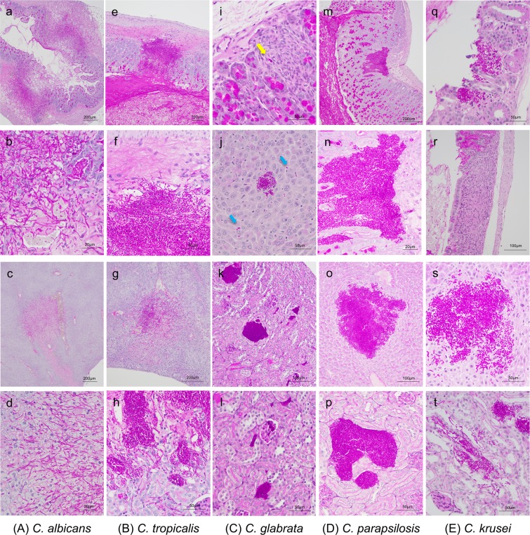
Histopathological examination using periodic acid-Schiff (PAS) staining. (a–d) Histopathological sections from DBA2/J mice infected with Candida albicans. C. albicans invaded into the gut mucosa and liver tissue resulting in marked necrosis. C. albicans was not identified in the kidney. (a) Cecum, magnification ×40; (b) cecum, magnification ×400; (c) liver, magnification ×40; or (d) liver, magnification ×400. (e–h) Histopathological sections from mice infected with Candida tropicalis. C. tropicalis invaded into the muscle layer of the colon. The majority of colonies were adjacent to blood vessels in the liver and were present in the renal pelvis and tubules in the kidney. (e) Colon, magnification ×100; (f) colon, magnification ×400; (g) liver, magnification ×100; and (h) kidney, magnification ×400. (i–l) Histopathological sections from mice infected with Candida glabrata. Only a few C. glabrata cells were observed in the gut (yellow arrow). C. glabrata was widely found in the liver along the sinusoid (blue arrows) with a few areas of accumulated yeast cells. The majority of colonies were located in the renal tubules and interstitium. (i) Colon, magnification ×400; (j) liver, magnification ×400; (k) kidney, magnification ×200; and (l) kidney, magnification ×400. (m–p) Histopathological sections from mice infected with Candida parapsilosis. C. parapsilosis cells were found in the gut mucosa in the colon; however, they did not invade into the muscularis mucosae. The majority of colonies were located in the renal pelvis and tubules in the kidneys. (m) Colon, magnification ×100; (n) colon, magnification ×400; (o) liver, magnification ×200; and (p) kidney, magnification ×400. (q–t) Histopathological sections from mice infected with Candida krusei. Only one location of accumulated C. krusei cells was detected in the colons of three mice (q). Several granulation tissues were observed in the colon (r). C. krusei presented as both yeast and hypha form in the liver and kidneys. (q) Colon, magnification ×100; (r) colon, magnification ×200; (s) liver, magnification ×400; and (t) kidney, magnification ×400.

