Fig. 2.
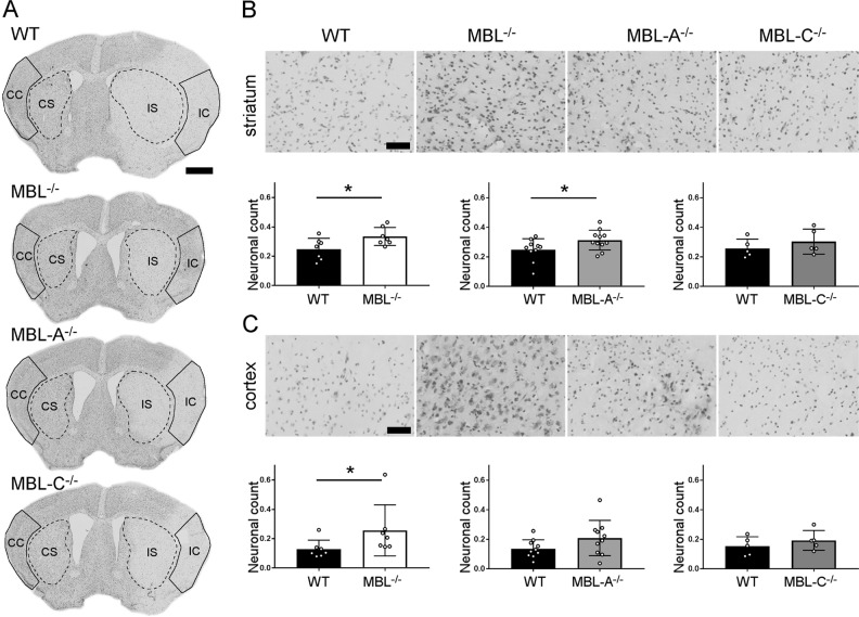
Neuronal count after 48 h of reperfusion in MBL−/−, MBL-A−/−, and MBL-C−/− mice. a Positioning of the cortical (outlines) and striatal (dotted outlines) regions of interest for calculating neuronal cell viability (CC: contralateral cortex; CS: contralateral striatum; IC: ipsilateral cortex; IS: ipsilateral striatum). The regions of interest were designed to include only the lesion area (pale cresyl violet staining) throughout the experimental groups. b, c Representative high-magnification images of cresyl violet-stained sections (scale bar, 50 µm) and neuronal counts showing that MBL−/− mice had more preserved neurons in the striatum and cortex and MBL-A−/− mice had more preserved neurons in the striatum than WT mice. The data are shown as bars with individual values ± SDs (n = 7 for MBL−/−, n = 10 for MBL-A−/−, and n = 5 for MBL-C−/−); t test, *p < 0.05. Compared with WT mice, MBL-C−/− mice showed no protection from ischemic injury

