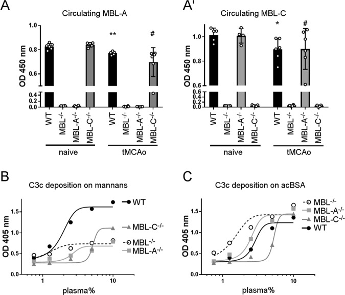
Fig. 5.
Plasma levels of MBL-A and MBL-C and systemic activation of the complement system after 48 h of reperfusion in MBL−/−, MBL-A−/−, and MBL-C−/− mice. a At 48 h after tMCAo, MBL-A was consumed to the same extent in MBL-C−/− mice as in WT mice (a). The data are shown as bars with individual values ± SDs (n = 5–6); Welch-corrected ANOVA for unequal variances, *p < 0.05 vs. naive WT mice, #p < 0.05 vs. naive MBL-C−/− mice. At 48 h after tMCAo, MBL-C was consumed to the same extent in MBL-A−/− mice as in WT mice (a'). The data are shown as bars with individual values ± SDs (n = 5–6); two-way ANOVA followed by Sidak’s post hoc test, *p < 0.05 vs. naive WT mice, #p < 0.05 vs. naive MBL-C−/− mice. b The in vitro assay for LP activation on mannans showed no activation in plasma from MBL−/− and MBL-A−/− mice but some activation in plasma from MBL-C−/− mice. c The in vitro assay for LP activation on acBSA showed similar activation in plasma from mice of each genotype. The data in b and c refer to pools of plasma from 5–6 mice per group, and plasma concentrations are reported on a logarithmic scale

