Fig. 1.
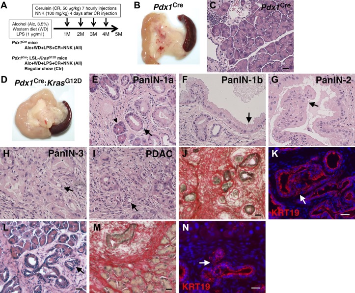
Promotion of pancreatic tumor development in Pdx1Cre;LSL-KrasG12D, but not in Pdx1Cre mice, by environmental risk factors. A: mouse models. Pdx1Cre mice and Pdx1Cre;LSL-KrasG12D mice were fed Western diet containing high cholesterol and high saturated fat, 3.5% alcohol, and LPS for 5 mo. Mice were also treated with cerulein and 4-(methylnitrosamino)-1-(3-pyridyl)-1-butanone (NNK) every month from 1st to 4th month. Pdx1Cre;LSL-KrasG12D mice were also fed a regular chow as a control (Ctr). B: gross appearance of the pancreas from Pdx1Cre mice treated with all insults for 5 mo. C: hematoxylin and eosin (H&E) staining of pancreas tissues from Pdx1Cre mice. D: gross appearance of the pancreas from Pdx1Cre;LSL-KrasG12D mice treated with all insults for 5 mo. E–K: H&E staining (E–I), Sirius red staining (J), and fluorescence immunostaining of cytokeratin 19 (KRT19; K) of pancreas tissues from Pdx1Cre;LSL-KrasG12D mice treated with all insults. L–N: H&E staining (L), Sirius red staining (M), and fluorescence immunostaining of KRT19 (N) of pancreas tissues from the control Pdx1Cre;LSL-KrasG12D mice fed a regular chow. An arrowhead indicates acinar cells undergoing acinar-ductal metaplasia. Arrows indicates pancreatic intraepithelial neoplasia (PanIN)-1a (E, L, and N), PanIN-1b (F), PanIN-2 (G), PanIN-3 (H and K), and pancreatic ductal adenocarcinoma (PDAC; I). Bar = 20 μm.

