Fig. 1.
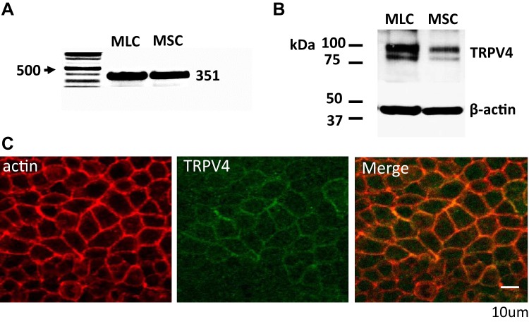
Expression and localization of transient receptor potential vanilloid member 4 (TRPV4) in mouse biliary epithelial cells. A: RT-PCR. TRPV4 primers were used to detect TRPV4 expression in both small (MSC) and large (MLC) mouse cholangiocytes, represented by a band size at 351 (see methods). Representative of 5 trials. B: Western blot utilizing polyclonal anti-TRPV4 antibody (see methods). TRPV4 protein (2 bands, at ~85 and ~100 kDa) is present in whole cell lysates of MLC and MSC cells; loading control was β-actin. Representative blot of 5 trials. C: membrane localization of TRPV4 protein in polarized MLC monolayers. Staining with anti-TRPV4 antibody (green) demonstrates TRPV4 protein localized with actin (red) in the plasma membrane. Scale bar, 10 μm.

