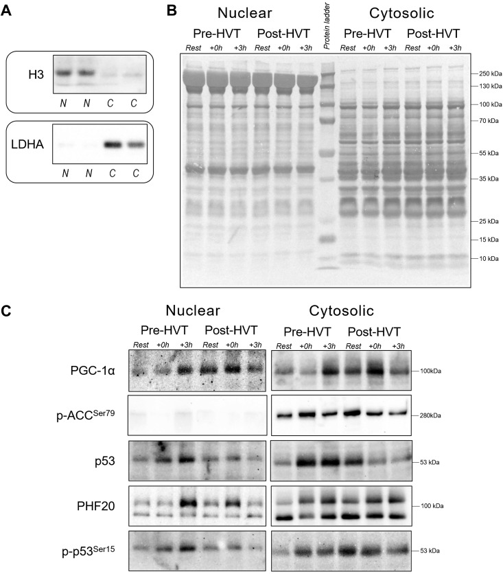
Fig. 2.
Subcellular enrichment, protein loading controls, and representative immunoblots. A: histone H3 and lactate dehydrogenase A (LDHA) were used as indicators of cytosolic (C) and nuclear (N) enrichment, respectively. B: whole lane Coomassie blue staining was used to verify equal loading between lanes in the nuclear and cytosolic fractions obtained from human vastus lateralis muscle biopsies before (rest), immediately postexercise (+0 h), and 3 h (+3 h) after a single session of high-intensity interval exercise (HIIE) performed at the same absolute intensity before (pre-HVT) and after (post-HVT) 40 sessions of twice-daily high-volume, high-intensity interval training (HVT). C: representative immunoblots of peroxisome proliferator-activated receptor γ coactivator-1α (PGC-1α), acetyl-CoA carboxylase phosphorylated at serine 79 (p-ACC Ser79), p53, plant homeodomain finger-containing protein 20 (PHF20; top band at 105 kDa), and p53 phosphorylated at serine 15 (p-p53 Ser15) measured in the same nuclear and cytosolic fractions. No band was detected in the nuclear fractions for p-ACC Ser79. The whole-lane Coomassie and immunoblot images in this figure were cropped to improve the conciseness and clarity of the presentation.

